Xiao Pan, Wenjing Liu, Chengtao Wang, Antian Gao, Zitong Lin
First published: 14 February 2025 https://doi.org/10.1111/iej.14209

Characteristics of vertical root fractures at early stages: Evidence that dentinal microcracks are an experimental phenomenon
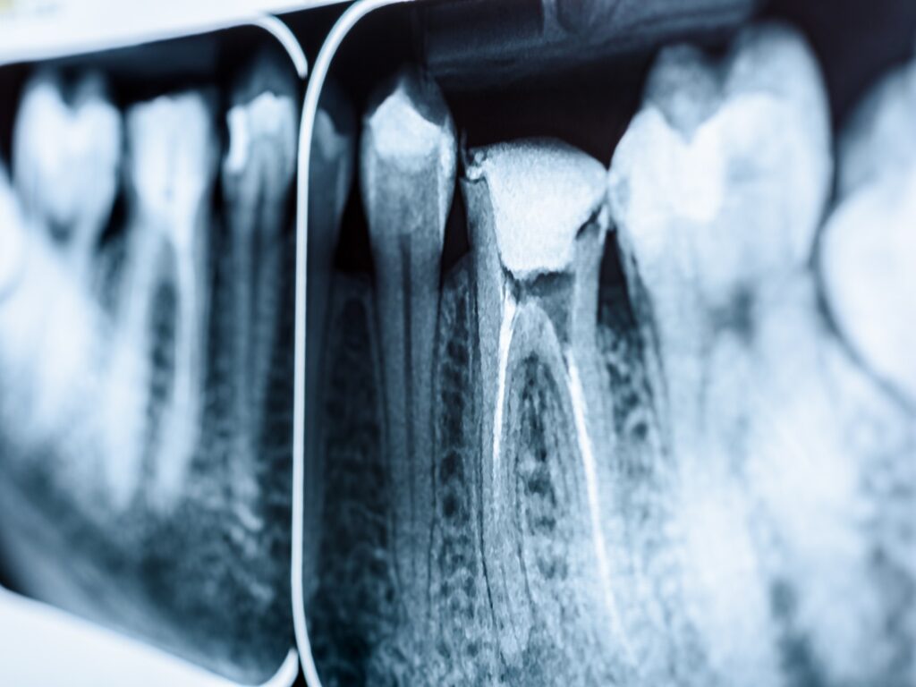
This study provides a refreshing and clinically relevant perspective on the early-stage morphology and pathogenesis of vertical root fractures (VRFs), challenging long-held assumptions that many of us in specialist practice have accepted for decades. Traditionally, the prevailing narrative has been that structural breakdown begins coronally, leading to pulpal involvement and eventual apical propagation (at least, that’s what I thought!). However, this paper, through the detailed micro-CT analysis of 28 early-stage VRF cases, presents compelling evidence that these fractures originate in the mid-root region, specifically at the canal wall, and propagate bucco-lingually toward the root surface. The meticulous imaging and analysis convincingly suggest that this internal-to-external crack trajectory—and not the surface-originating microcracks described in numerous earlier in vitro studies—is the true morphological hallmark of early VRFs.
One of the most thought-provoking implications of this paper lies in its critique of the concept of dentinal microcracks as precursors to VRFs. The authors argue that many such microcracks previously reported are artefacts—introduced during extraction, sectioning, or dehydration in lab settings—and are not a reliable representation of in vivo fracture initiation. The consistent bucco-lingual direction, mid-root origin, and root canal wall involvement in all observed fractures stand in contrast to the variable and often superficial nature of microcracks reported in prior research. For clinicians, especially those using CBCT for diagnosis, these findings are valuable: even in high-resolution CBCT, fractures of <150 μm width—like those found here—will elude detection. I would have thought that a lateral lesion without extension to crestal bone would most likely be due to a lateral canal. Here is proof that it is very likely to be an early VRF.
While the study is well-conceived and rigorously executed, it is not without limitations. The sample size, though meaningful given the rarity of verifiable early-stage VRFs, remains small and geographically restricted. Furthermore, despite best efforts, the possibility of microcrack formation during extraction, even by experienced surgeons, cannot be entirely dismissed. It would also have been valuable to see a control group of endodontically treated but non-fractured teeth scanned under identical conditions to further substantiate the claim that the observed crack morphologies are truly pathognomonic. Nonetheless, this paper is an important contribution to our understanding of VRFs, and its insights should influence both diagnostic vigilance and future research into fracture mechanics in endodontically treated teeth.