There are certain moments in dentistry when time appears to stand still.
You are looking for a canal. Perhaps the chamber is heavily calcified. Perhaps the tooth is rotated. Perhaps the crown bears only a passing resemblance to the tooth underneath it.
You extend the access a little further.
And then the bur drops.
There is bleeding.
Quite a lot of bleeding.
You stare down the access cavity and somewhere in the back of your mind a small voice says:
“That’s not the canal.”
This is the moment you need Winston Wolf.
First rule: don’t panic
A perforation is an artificial communication between the root canal system and the periodontal tissues. It may occur during access preparation, while searching for a canal, during instrumentation or during post preparation. It can also occur pathologically, for example as a consequence of resorption.
But for the purposes of today, we are talking about the one you just made.
The good news is that a perforation does not necessarily condemn the tooth. Modern magnification, imaging and hydraulic calcium-silicate materials mean that many perforations can be managed successfully.
The bad news is that what you do after making the perforation can dramatically change the prognosis.
So, before reaching for another bur, file or bottle of irrigant, remember Winston’s advice:
STOP
S — Stop
T — Take stock
O — Obtain information
P — Protect the tissues
Let’s clean this mess up properly.
This sounds ridiculously obvious.
It isn’t.
One of the most dangerous moments is the period immediately after the operator suspects that something has gone wrong.
The natural temptation is to investigate.
Perhaps if I just extend a little further…
Perhaps the canal is underneath this…
Perhaps that bleeding point actually is the canal…
Stop.
If you are drilling in the wrong place, additional drilling rarely improves matters.
The same principle applies during instrumentation. If a file suddenly appears to be somewhere unexpected on a working-length radiograph, resist the temptation to continue enlarging the “canal” until you understand where the instrument actually is.
A small perforation is generally easier to manage than a large one.
Your first objective, therefore, is remarkably simple:
Do not turn a small problem into a big one.
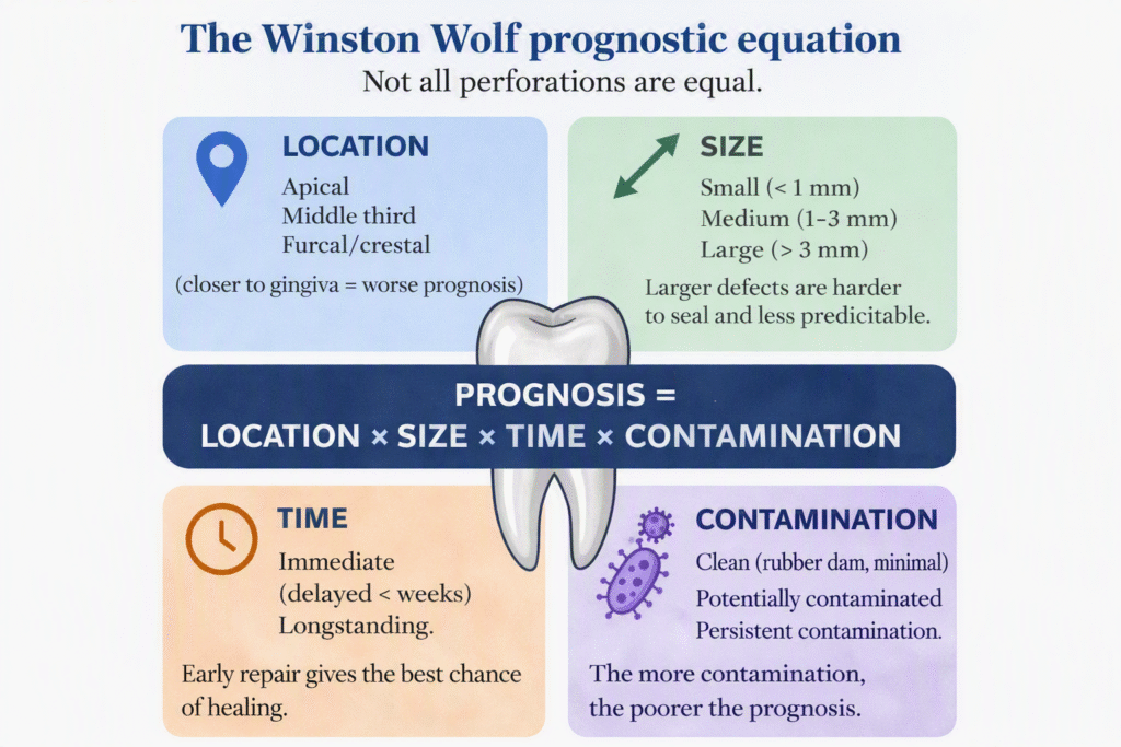
Not all perforations are created equal.
Before deciding what to do, there are four questions Winston wants answered:
WHERE IS IT?
A small apical perforation is very different from a furcal perforation close to the epithelial attachment.
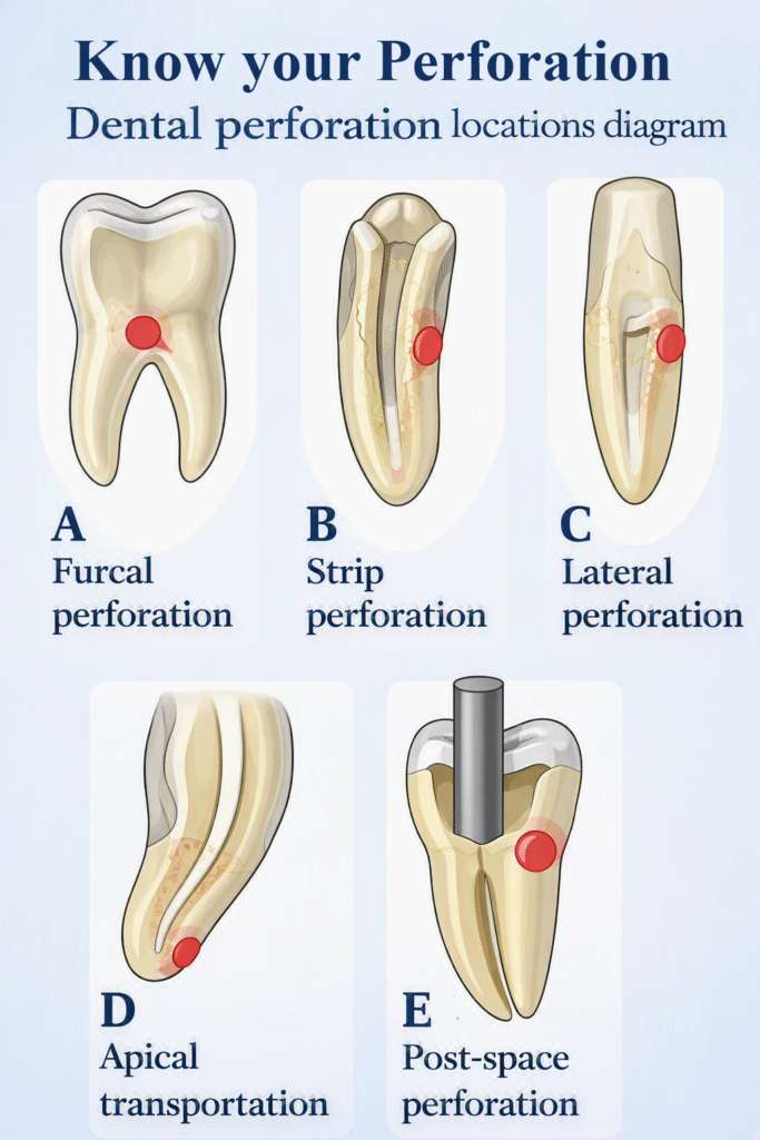
Broadly, perforations may occur:
Location matters because a perforation close to the crestal bone and gingival attachment is at greater risk of developing a periodontal communication.
Once that communication becomes established, the biological problem becomes considerably more difficult.
HOW BIG IS IT?
Size matters.
A tiny perforation produced by the tip of an ultrasonic instrument is a very different proposition from a large defect created with a high-speed bur.
Apart from the obvious increase in tissue injury, larger defects are harder to seal predictably and make extrusion of repair material more difficult to control.
WHEN DID IT HAPPEN?
If the answer is:
“About thirty seconds ago”
…that is generally good news.
If the answer is:
“I think it may have happened during the root canal treatment six months ago”
…Winston is considerably less cheerful.
An immediately recognised perforation under rubber dam has had limited opportunity for bacterial contamination. A longstanding perforation may already be associated with inflammation, periodontal breakdown and bone loss.
WHAT HAS BEEN THROUGH IT?
This is sometimes forgotten.
A fresh mechanical perforation and an infected perforation are not biologically equivalent.
Has the defect been exposed to saliva?
Has it been open beneath a temporary restoration?
Is there already a periodontal pocket communicating with it?
And, importantly, what have you put through it since it happened?
Which brings us to an important Winston Wolf principle:
If you don’t know where a hole goes, don’t pour sodium hypochlorite into it.
If you remember nothing else from this article, remember these four factors:
LOCATION × SIZE × TIME × CONTAMINATION
They are not genuinely a mathematical equation, of course, but together they provide a useful way of thinking about prognosis.
A small, freshly created, immediately recognised perforation away from the gingival attachment may have a very reasonable prognosis.
A large, longstanding perforation associated with periodontal communication and osseous destruction is an entirely different animal.
This distinction is important when talking to patients.
The word perforation sounds catastrophic.
Sometimes it is.
Often it isn’t.
The correct question is not simply:
“Is there a perforation?”
It is:
“What sort of perforation are we dealing with?”
Now we need to establish exactly what has happened.
Magnification
If you have a dental operating microscope, this is the time to use it.
Clean the chamber carefully, obtain haemostasis and inspect the anatomy.
Can you see the perforation?
Can you identify the true canal orifices?
Can you distinguish the chamber floor from the repair site?
The answer to those questions determines what happens next.
Radiographs
Take an appropriate periapical radiograph and consider changing the horizontal angulation.
If an instrument has inadvertently passed through a perforation, an angled radiograph may help determine whether it is following the expected root anatomy.
CBCT
In selected cases, a small-volume CBCT can be extremely helpful.
It can help determine the location and extent of the defect, its relationship to the crestal bone and periodontal tissues, and—critically—where the actual canal is.
But CBCT is not a substitute for thinking.
The question is not:
“Can I take a CBCT?”
It is:
“Will a CBCT give me information that changes what I do next?”
If it will, take one.


Preop CBCT showing furcal perforation with associated radiolucency
Clinical image showing bleeding perforation site
Clinical image showing perforation repair with boiceramic putty, following location of the true canal
Comparative review CBCT slices showing complete bony healing of the previous furcal radiolucency
Three-dimensional imaging can be invaluable when conventional imaging does not adequately reveal the relationship between the perforation, true canal and periodontal tissues.
Once you know what you are dealing with, the objective is to create conditions in which the tissues have the best opportunity to heal.
That means controlling contamination, achieving haemostasis and sealing the communication with an appropriate biocompatible material.
Modern hydraulic calcium-silicate materials have transformed our ability to manage these defects.
MTA established the biological principles and remains supported by substantial clinical evidence. Newer calcium-silicate materials may offer handling, setting-time and discolouration advantages depending on the clinical situation.
But here is the important point:
The material is not the treatment.
A beautifully placed blob of bioceramic material does not compensate for:
Successful perforation management begins before the repair material comes out of the packet.
Perforations tend to present in several recognisable ways.
The classic access catastrophe.
The operator searches for a canal, loses orientation and gradually heads towards the furcation.
These can be particularly problematic because of their proximity to the periodontal attachment.
The earlier the problem is recognised and sealed, the better.
Usually the result of excessive removal of dentine from the inner wall of a curved root.
The mesial root of a mandibular molar is the classic example.
This is why understanding the concept of the danger zone matters. We want to shape canals while preserving strategically important radicular dentine—not produce the world’s most impressive taper at the expense of the root.
Often encountered while searching for calcified or displaced canals.
This is where the external shape of the crown can deceive you.
Crowns, restorations, rotation, tilting and secondary dentine can all distort the landmarks we normally rely upon.
The tooth does not care where you think the canal ought to be.
Sometimes we reach the correct working length.
Unfortunately, we have reached it by creating our own route.
Loss of the original canal trajectory can lead to transportation, ledging and ultimately perforation.
Prevention starts with appreciating anatomy and curvature before instrumentation begins.
A slightly different crime scene, but the same victim.
When preparing a post space, losing the long axis of the root can result in lateral perforation. Existing crowns and bridges can make orientation particularly difficult.
As always, prevention is considerably easier than repair.
Once a perforation has occurred, there are several approaches Winston would advise against.
Winston does not keep drilling.
If you have lost orientation, stop and re-establish it.
Winston does not irrigate enthusiastically through an unidentified bleeding hole.
Sodium hypochlorite is an excellent endodontic irrigant.
The periodontal tissues are less enthusiastic about it.
Winston does not attempt to repair something he cannot see.
Blindly pushing repair material towards a perforation is not precision endodontics.
Winston does not fill the defect with whatever happens to be closest on the materials trolley.
This is a biological repair. Use a material designed for the job.
Winston does not forget about the actual root canal.
Congratulations—you repaired the perforation.
There is still an infected root canal system to clean, shape and obturate.
And Winston does not tell the patient:
“Everything went absolutely fine.”
If you have caused a perforation, tell the patient.
Explain what happened in language they can understand.
Explain what you propose to do about it.
Explain whether it changes the prognosis.
And document the conversation.
Trying to hide a complication is not only professionally problematic; it also destroys trust if the patient subsequently discovers what happened.
Complications occur.
What defines us professionally is how we deal with them.
There is a considerable difference between saying:
“Unfortunately, during treatment an opening has occurred between the inside of the tooth and the surrounding tissues. I’ve recognised it immediately. These can often be repaired, but it changes the complexity of the treatment and I’d like to discuss the options with you.”
…and hoping nobody notices.
Patients are generally much more understanding of complications than they are of surprises.
Perhaps.
The important question is not whether a general dentist can repair a perforation.
It is whether this particular perforation, in this particular tooth, with your available equipment and experience, is one you can manage predictably.
Ask yourself:
Can I see it?
Can I control the bleeding?
Can I identify the true canal?
Can I place the repair material accurately?
Can I assess the periodontal implications?
Can I complete the remainder of the endodontic treatment predictably?
If the answer to several of those questions is no, referral is not failure.
Referral is treatment planning.
And there is an important timing issue here.
Refer before attempting three different repair techniques.
A small, fresh perforation is usually a much nicer referral than a large perforation containing amalgam, glass ionomer, temporary filling material and several optimistic layers of MTA.
Winston prefers arriving at the crime scene before everybody has walked through the evidence.
The best perforation repair remains prevention.
Before starting treatment, look carefully at:
Do not assume that the long axis of the crown is the long axis of the root.
Once you enter the pulp chamber, use anatomical landmarks rather than simply drilling to an arbitrary depth.
In calcified cases, slow down.
Magnification and illumination can transform the procedure. Ultrasonics allow controlled dentine removal. In carefully selected cases, CBCT can provide information that conventional radiography cannot.
And when calcification is severe, guided endodontic access, static or dynamic may allow us to reach canals while preserving dentine and reducing the risk of losing orientation.
Technology does not replace clinical judgement.
It gives clinical judgement better information.

So, the next time the bur drops and you see rather more blood than you were expecting:
STOP.
Stop before making the defect larger.
Take stock of its location, size, timing and likely contamination.
Obtain information using magnification, appropriate radiography and CBCT where indicated.
Protect the tissues by controlling contamination and sealing the defect appropriately.
And remember:
A perforation isn’t necessarily the end of the tooth.
With early recognition, careful assessment and appropriate management, many perforated teeth can still be retained successfully.
But if you suddenly find yourself somewhere you weren’t planning to be, Winston has one final piece of advice:
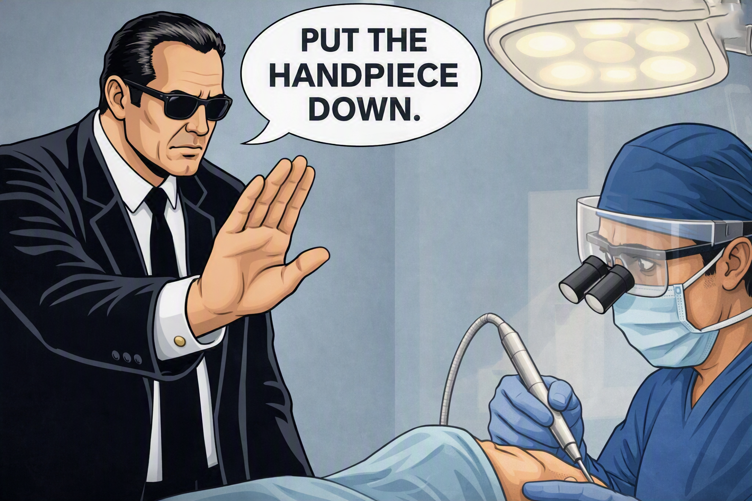
Put the handpiece down.
It is much easier to solve one problem than two.
Suggested key references
Our dentists and nurses are all registered with the General Dental Council
The practice is regulated by the Care Quality Commission
Our complaints process can be found here.



© 2026 Academy of Advanced Endodontics. All rights reserved.